3D Imaging/Cone Beam
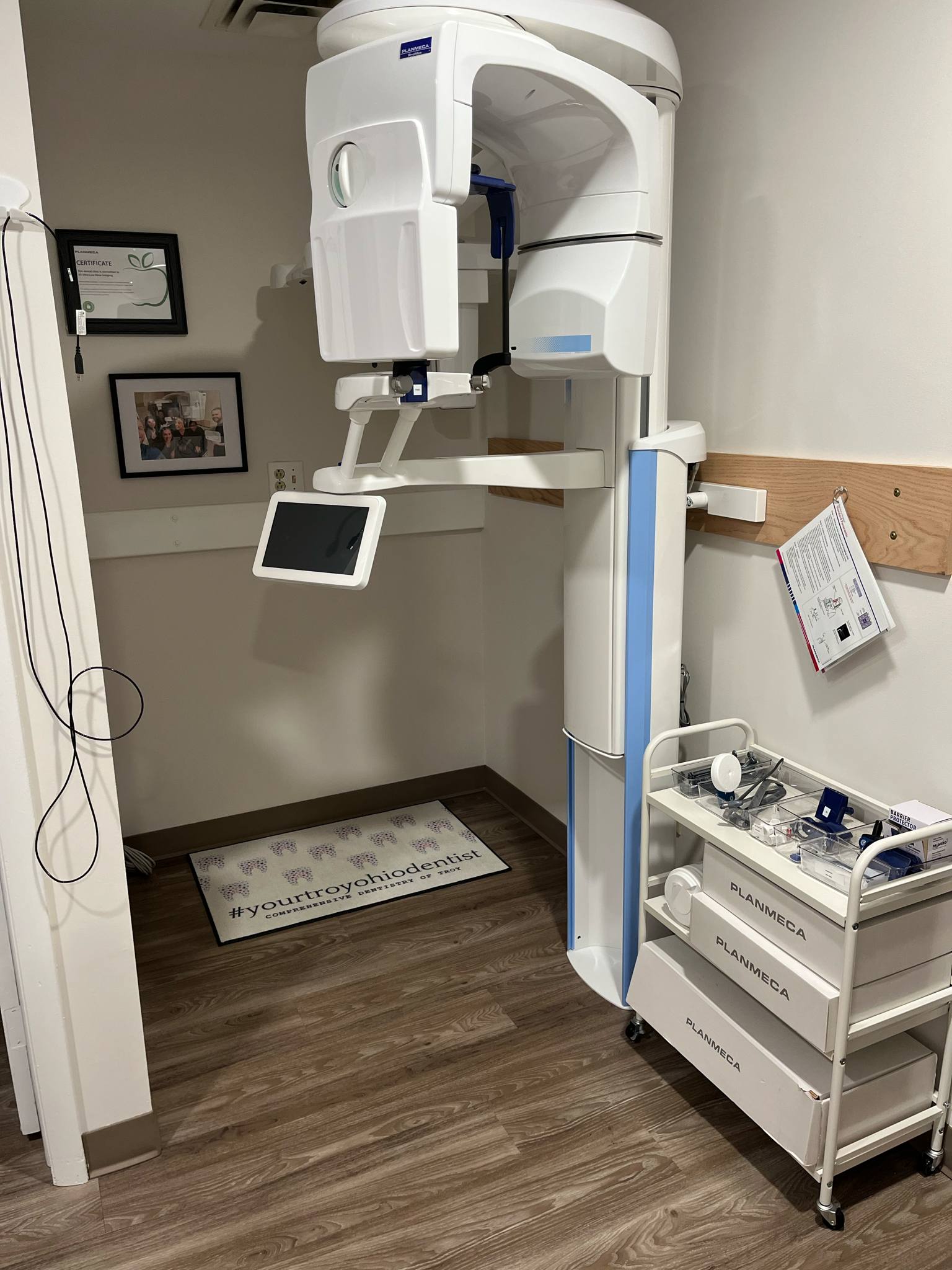
CBCT (Cone Beam Computed Tomography) is a specialized X-ray imaging technique used in dentistry to produce 3D images of the teeth, jaws, and surrounding tissues. It provides a more detailed view than traditional 2D dental X-rays, enabling dentists to better visualize and diagnose dental conditions and plan treatments, especially for procedures like dental implants. Key features and benefits of CBCT: 3D Imaging: CBCT captures detailed 3D images, allowing dentists to visualize the anatomy of the teeth, jaws, and surrounding structures from multiple angles. Accurate Diagnosis: The detailed 3D images facilitate more accurate diagnosis of dental conditions like impacted teeth, cysts, and fractures, according to the American Association of Endodontists. Improved Treatment Planning: CBCT images help dentists plan complex procedures like dental implants, root canals, and orthodontic treatment, ensuring precise placement and reducing the risk of complications, according to the U.S. Food and Drug Administration. Reduced Radiation Exposure: Compared to conventional CT scans, CBCT exposes patients to significantly less radiation, according to Maven Imaging. Faster Scan Times: CBCT scans are generally faster than conventional CT scans, requiring less time for the patient to remain still.
Referring Providers: Please click the link to view and complete the required online referral: